Anterior blepharitis, meibomian gland dysfunction and dry eye disease are all common conditions that, if left untreated, can thwart postoperative success. Optometrists actively manage these prior to surgery. But what about other corneal conditions that can affect refractive outcomes, such as pterygia, corneal guttata, Fuchs’ dystrophy, epithelial basement membrane dystrophy and superficial corneal opacities? These can also undermine surgical outcomes due to poor tear distribution.
 |
 |
|
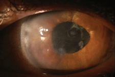
| Salzmann's nodular dystrophy.
|
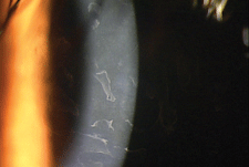
Anterior basement membrane dystrophy.
|
Many surgeons commonly perform superficial keratectomy prior to cataract surgery to prevent adverse outcomes. Referring ODs should be attuned to the scenarios in which this is warranted and make the appropriate recommendation. Three of note are as follows:
• Salzmann’s nodular degeneration presents with bluish-white peripheral nodules raised above the corneal surface. It is slowly progressive, more prevalent in females and often occurs in tandem with long-standing scars and chronic uveitis. The raised surface may be associated with tear film abnormalities, irregular astigmatism and difficulties in contact lens fitting.1
• Band keratopathy is characterized by the appearance of a band across the central cornea, formed by the precipitation of calcium salts on the corneal surface (directly under the epithelium).2 Its etiology is typically previous infection or inflammation. Depending on depth of corneal penetration, superficial opacities can be removed. Visual acuity can be impaired depending on the density of the corneal salts.
• Anterior basement membrane dystrophy is the most common anterior corneal dystrophy seen in practice. Clinical findings include bilateral presentations, map-like patterns, fingerprint lines, fine dots (microcysts) or comma-shaped opacities that may be variable over time. It can lead to recurrent corneal erosion and blurred or double vision due to the poor adhesion to the corneal basement membrane.
Depending on surgeon preference, superficial keratectomy can be performed in the operating room or at the slit lamp. Topical proparacaine or tetracaine is used for anesthesia. Prior to removal of the opacity, sodium fluorescein is used to identify surface irregularities and “sick” epithelium. Using a dry cellulose sponge or a scarifier blade, the treatment is limited to the corneal epithelium, which is removed down to Bowman’s layer. After removal, the clear corneal surface is polished with a cellulose sponge, topical antibiotics are administered, and a bandage lens is placed to promote healing and patient comfort.1
Post-op care is similar to photorefractive keratectomy, with visits at one day, one week and one month. Patients are prescribed topical antibiotics and NSAIDs QID for one week, and topical steroids over several weeks to minimize corneal haze and scarring. Complications include a recurrence of corneal condition, infection, HSV reactivation and delayed epithelial healing. When the cornea is healed in one to two months, patients can be reappointed for cataract evaluation.
1. Available at: http://oculist.net/downaton502/prof/ebook/duanes/pages/v6/v6c028.html.2. Available at: http://emedicine.medscape.com/article/1194813-overview.

