 |
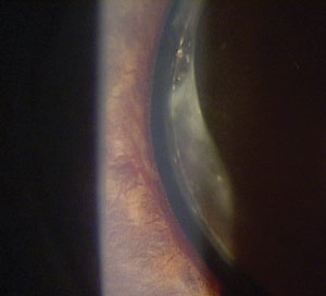
This study demonstrated that the presence of a PVD is protective against development of NVG in eyes with prior retinal vascular occlusion. Photo: Lee Peplinski, OD. Click image to enlarge. |
New data uncovered a number of factors associated with an increased risk of developing neovascular glaucoma (NVG) after retinal vascular occlusion, including Black race, Hispanic/Latino ethnicity, central retinal vein occlusion (CRVO), diabetic retinopathy and the absence of posterior vitreous detachment.
Recently published in the journal Ophthalmology and Therapy, this research explored the connection between posterior vitreous detachment (PVD) and other potential factors with the risk of developing neovascular glaucoma in eyes affected by occlusions of the retinal artery (RAO) or retinal vein (RVO).
This single-center, retrospective, case-control study included adults with a history of RVO/RAO. Patients who developed neovascular glaucoma (n=101) were age- and sex-matched 1:2 to controls who did not develop NVG (n=202).
Initial analyses showed no difference in risk of neovascular glaucoma based on eye, lens status, hypertension, history of panretinal photocoagulation or retinal surgery. However, a borderline difference was observed based on diabetic retinopathy (DR) and prior anti-VEGF treatment. Researchers reported a significant difference based on race/ethnicity, type of vascular event and PVD status.
The final model revealed that patients without PVD were significantly more likely to develop neovascular glaucoma independent of other covariates, according to the study authors. “In this case-control study, several factors were independently associated with higher likelihood of developing neovascular glaucoma following a retinal vein or artery occlusion: absence of a posterior vitreous detachment, Hispanic/Latino ethnicity, Black race, CRVO and diabetic retinopathy,” the study authors noted in their recent Ophthalmology and Therapy paper. “Thus, presence of a posterior vitreous detachment was protective against neovascular glaucoma, which may suggest some relationship between the vitreous interface and NVG risk,” they noted in the paper.
“Patients with one or more of these characteristics at the time of presentation for a vascular occlusion may benefit from more frequent monitoring for neovascularization of the angle and intraocular pressure trends,” they concluded. “Whether such patients may benefit from more aggressive prophylactic interventions should be considered in future studies.”
| Click here for journal source. |
Palmer LD, Peterson JD, Evans JK, et al. Posterior vitreous detachment and risk of neovascular glaucoma in eyes with prior retinal vascular occlusions. Ophthalmol Ther. September 29, 2024 [Epub ahead of print]. |

