A recent study, which used OCT angiography (OCT-A) to evaluate type 1 and type 2 macular neovascularization (MNV) components in age-related macular degeneration (AMD), found that type 1 and type 2 MNV may respond differently to anti-VEGF treatment over the course of one year.
This retrospective analysis involved eyes with treatment-naïve exudative AMD treated with anti-VEGF injections under a PRN protocol over one year. Two-dimensional areas and three-dimensional volumes of MNV were obtained from macular PR-OCTA scans using an automated convolutional neural network. Macular neovascularization was identified as a flow signal within the outer retinal slab, with type 1 and type 2 components analyzed individually.
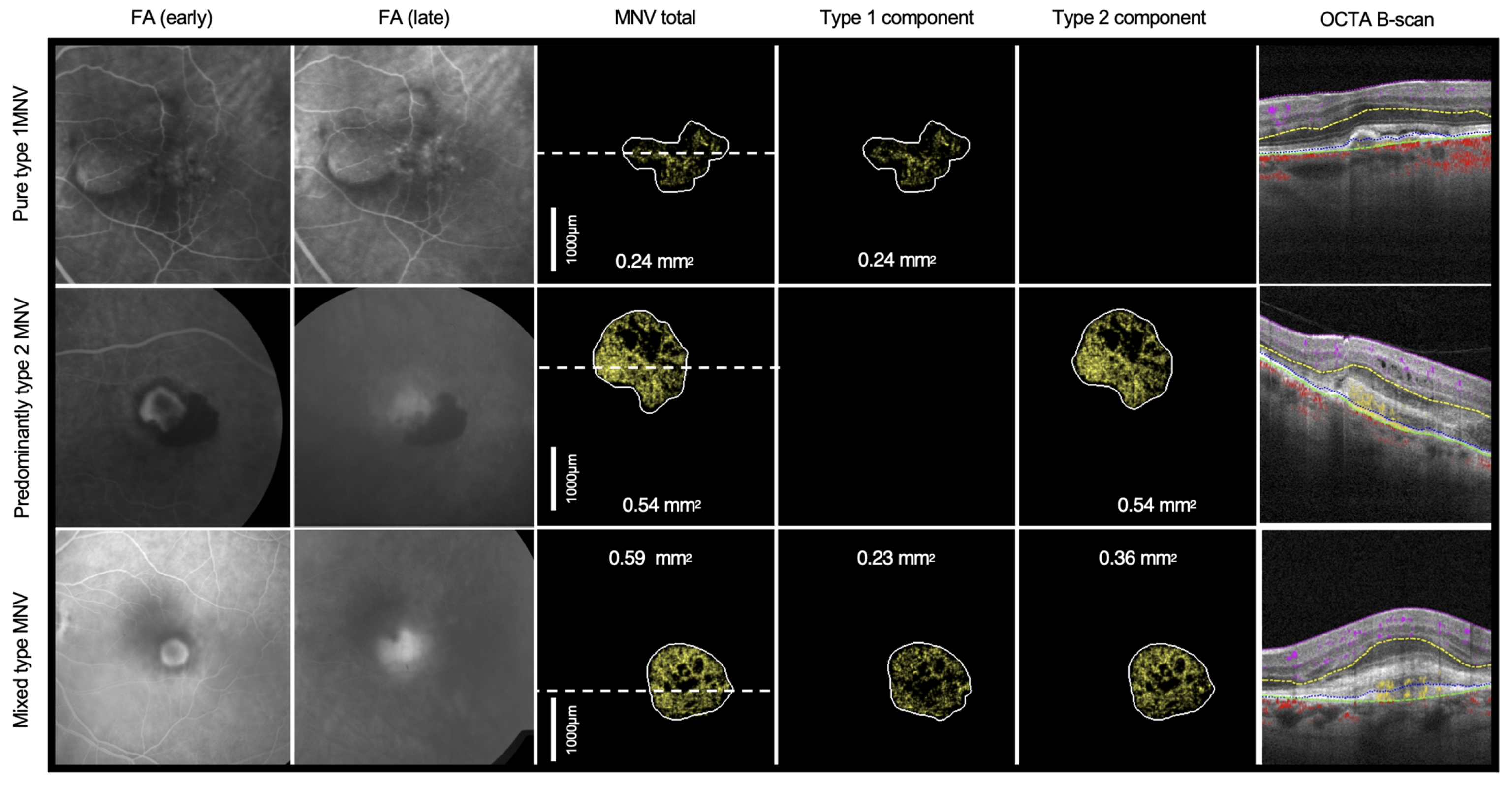 |
| OCT-A scans reveal distinct responses in type 1 and type 2 macular neovascularization following one year of anti-VEGF treatment in patients with age-related macular degeneration. This image from the study shows representative examples of MNV type based on OCT-A classification, with corresponding early and late FA. Photo: Tsuboi K, et al. Invest Ophthalmol Vis Sci. 2024;65(11):32. Click image to enlarge. |
Among the 17 enrolled eyes, 12 were pure type 1 MNV and five were type 2 MNV. In eyes with pure type 1, the study authors found that the total—sum of type 1 and type 2 components—MNV area and volume did not change from baseline to six months or 12 months. Comparatively, a significant decrease in total macular neovascularization area from baseline to six months and 12 months was observed among eyes with type 2 MNV.
“The total type 2 MNV volume also decreased from baseline visit to visits at six months and at 12 months, nearing statistical significance,” the research team reported. “In eyes with type 2 MNV, the type 1 component increased from 0.093mm2 to 0.30mm2, and the type 2 component decreased from 0.37mm2 at six months to zero at 12 months.”
While summarizing their research in the journal Investigative Ophthalmology & Visual Science, the study authors noted, “we provide evidence suggesting that type 2 MNV has greater reductions in MNV area and MNV volume compared to type MNV at six months and one year under PRN anti-VEGF treatment.
“Although the current small study will not change current clinical management, it does demonstrate the ability to automatically quantify type 1 and type 2 MNV using OCT-A,” they concluded. “Further studies with larger sample sizes, different treatment protocols, longer follow-up and different anti-VEGF agents may reveal useful clinical management information, provide prognostic clues and improve our understanding of MNV pathogenesis.”
| Click here for journal source. |
Tsuboi K, You QS, Wang J, et al. Quantitative Evaluation of Type 1 and Type 2 Choroidal Neovascularization Components Under Treatment With Projection-Resolved OCT Angiography. Invest Ophthalmol Vis Sci. 2024;65(11):32. |