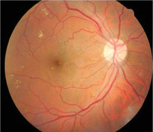
 Researchers at UC Berkeley School of Optometry have found that digital photos can help detect hard exudates—a key early sign of diabetes-related macular edema. Their findings were published in the April issue of Optometry and Vision Science.
Macular edema is one of the most threatening visual conditions faced by people with diabetes. Unfortunately, about 20% of patients already have early signs of the condition when they’re diagnosed, and many aren’t under the continued care of an eye care professional.
The researchers took magnified retinal images of 103 adults with type 2 diabetes who were considered at risk for macular edema. Researchers did not use drops to dilate the eye and took the images at a public health clinic using the EyePACS teleophthalmology system. The photos were then sent to eye specialists online, who looked for hard exudates close to the line of sight as an indicator of clinically significant macular edema.
Follow-up dilated exams by eye specialists showed clinically significant macular edema in about 15% of patients, suggesting that hard exudates detected on the digital photographs were an accurate indicator of macular edema.
“Since the completion of that study, we have looked at whether the presence of hard exudates within 500µm of the fovea is associated with greater retinal thickness measured by OCT,” says lead author Taras Litvin, OD. “We found that patients had greater central macular thickness on the OCT thickness maps if they had exudates less than 500µm from the fovea on OCT scans.”
The presence of hard exudates located within one disc diameter of the foveola in diabetes patients is a reliable marker for clinically significant macular edema. This diagnostic method, however, shouldn’t replace a dilated fundus exam, which includes careful biomicroscopy when such an exam is possible, Dr. Litvin added.
Litvin TV, Ozawa GY, Bresnick GH, et al. Utility of hard exudates for the screening of macular edema. Optom Vis Sci. 2014 Apr;91(4):370-5.

