 |
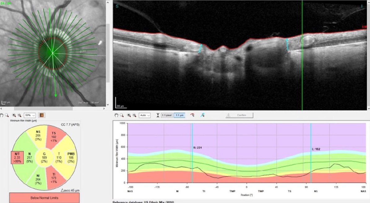
A computer simulation-based approach investigated risk factors for POAG progression. In the early stages of POAG, the rate of decrease in RNFL thickness accelerated with increasing mean or maximum strain on the lamina cribrosa. Additionally, the lamina cribrosa strain was associated with ONH geometric factors and IOP. Photo: James L. Fanelli, OD. Click image to enlarge. |
Many have suggested that the optic nerve head (ONH) strain response to intraocular pressure (IOP) may be a useful novel biomarker for predicting vulnerability to glaucomatous damage. However, performing ONH strain mapping on OCT imaging using recent technology appears to be challenging because the strain is highly sensitive to tissue composition and topography. Researchers in Korea believed that computer simulation is useful for predicting biomechanical stress and strain in the ONH. They recently conducted a computer simulation of the ONH to evaluate the factors related to the ONH strain and stress and to investigate the risk factors associated with the progression of primary open-angle glaucoma (POAG).
They found that, in the early stages of POAG, the rate of decrease in retinal nerve fiber layer (RNFL) thickness accelerated with increasing mean or maximum strain on the lamina cribrosa. Additionally, the lamina cribrosa strain was associated with ONH geometric factors and IOP.
This retrospective analysis, which was published in Journal of Glaucoma, included patients diagnosed with early-to-moderate stage POAG. The strains and stresses on the RNFL surface, prelaminar region, and lamina cribrosa were calculated using computer simulations based on finite element analysis. The correlations between the rate of RNFL thickness decrease and biomechanical stress and strain were investigated in both the progression (n=71) and nonprogression groups (n=47).
In the progression group, negative correlations with the RNFL thickness decrease rate included the maximum and mean strain on the lamina cribrosa. In multivariate analysis, the mean strain on the lamina cribrosa was associated with optic disc radius, optic cup deepening, axial length and mean IOP, whereas the maximum strain was only associated with mean IOP.
The simulation revealed that: (1) the lamina cribrosa, rather than the prelaminar tissue, was identified as a region associated with the progression; (2) no correlation was observed between the rate of RNFL thickness decrease and mechanical strain or stress on ONH in the non-progression group; and (3) a significant association was observed between ONH geometric parameters and the mean strain on the LC.
“Our findings suggest that IOP reduction is important and ONH geometric factors may be considered as risk factors for progression,” the study authors wrote in their paper.
They suggested that the investigation of lamina cribrosa strain resulting from longitudinal changes in optic cup deepening is needed for providing comprehension of the association of optic cup deepening with the glaucoma progression.
“Computer-aided simulations of glaucoma can offer insights into the management and understanding of the pathophysiology of its development and progression,” the researchers highlighted. “If automated modeling and simulation technologies are enhanced in the future, they may be employed to predict glaucoma progression in clinical settings.”
They concluded that these findings underscore the importance of IOP reduction in the early stages of glaucoma management. Moreover, their results suggest that optic disc geometry could potentially serve as a biomarker of progression.
| Click here for journal source. |
Kang E, Park JH, Yoo C, Kim YY. Effects of stress and strain on the optic nerve head on the progression of glaucoma. J Glaucoma. October 7, 2024. [Epub ahead of print]. |

