 |
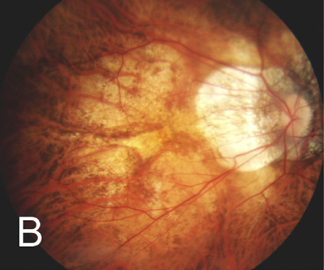
Regional and ethnic differences in axial elongation are age-specific, a recent study determined. These two demographic factors had the greatest effect in younger eyes with myopia, although region and ethnicity also appeared to affect axial elongation estimates in emmetropic eyes. Photo: Julie Poteet, OD. Click image to enlarge. |
Researchers have been studying how various demographic factors influence axial growth patterns, which can help develop growth charts to aid myopia management. Three of these factors include age, region and ethnicity, all of which have been shown to affect axial elongation and myopia progression; however, debate remains surrounding the extent of these differences. Aiming to improve the availability of normative progression data, a new retrospective analysis evaluated axial elongation and spherical equivalent (SE) progression in non-myopic and myopic children from Asian and European regions. They drew several conclusions from their findings, published in Ophthalmic and Physiological Optics, which we dive into below.
The study leveraged data from 15 longitudinal clinical and population-based studies distributed across the UK, Sweden, Australia (Europe), China and Vietnam (East Asia). Researchers included 14,593 data points from 6,208 participants aged six to 16 years with an SE range from +6D to -6D. Axial elongation and cycloplegic SE refraction were annualized from these studies.
The results indicated that axial elongation and SE progression were generally lower in children from European regions than those in East Asia. The magnitude of these regional/ethnic differences depended on myopia status and age, with larger differences observed in myopes (compared with non-myopes) and in patients of younger ages.
“Age-specific regional/ethnic differences indicated that axial elongation for a six-year-old East Asian myopic child was greater than a European child by 0.15mm/year (0.58 vs. 0.43mm/year) and by 0.09mm/ year (0.35 vs. 0.26mm/year) for a 10-year-old myope,” the researchers reported in their paper on the study. Additionally, they noted, “SE progression was lower in a six-year-old European myope by 0.48D/year and at 10 years of age by 0.34D/year compared with an East Asian myope.”
In the discussion portion of their paper, the researchers speculated about why regional/ethnic differences were greater in children of younger ages, which prior studies have also observed. They explained that this finding could be related to “the greater exposure to education and emphasis on reading prior to starting formal schooling in the East/Southeast Asian region, where myopia is associated with higher educational achievements.” These observations, they noted, “highlight the need to identify individuals at risk of progression and commence preventive and therapeutic myopia management at an early age and monitor progression using age-specific tools.”
Another interesting conclusion of this study is that regional/ethnic differences in axial elongation present not only in myopes, but also in non-myopes. This conflicts with a previous study, which noted that ethnic differences in axial elongation were not significant in persistent emmetropic eyes. The researchers explained that because the current study defined non-myopes after only one year of observation, “it is possible that our sample of persistent non-myopes included eyes that would become myopic after one year, which is more likely in the Asian region.” Thus, they suggested, “the present findings highlight the need and the means to identify and monitor high-progressing non-myopic children.”
In summary, the authors note that these population-based estimates of axial elongation and myopia progression reveal substantial regional and ethnic differences in both myopic and non-myopic children that are reduced with increasing age. “Model-based estimates by age, gender and region/ethnicity may be used to assess myopia treatment efficacy,” they concluded.
| Click here for journal source. |
Naduvilath T, He X, Saunders K, et al. Regional/ethnic differences in ocular axial elongation and refractive error progression in myopic and non-myopic children. Ophthalmic Physiol Opt. 2024;1-171. |

